Pleomorphic Xanthoastrocytoma : A Pleomorphic Xanthoastrocytoma Who Grade Ii Pleomorphic Download Scientific Diagram : Pxa was first reported by kepes et al,1 and because of its proclivity for the temporal lobes, commonly presents with seizures.
Pleomorphic Xanthoastrocytoma : A Pleomorphic Xanthoastrocytoma Who Grade Ii Pleomorphic Download Scientific Diagram : Pxa was first reported by kepes et al,1 and because of its proclivity for the temporal lobes, commonly presents with seizures.. Composite pleomorphic xanthoastrocytoma and ganglioglioma: Pleomorphic xanthoastrocytoma treatment found 3 result. Patients have shorter survival rates when compared to those with who grade ii pleomorphic xanthoastrocytoma. Case report and literature review. Pleomorphic xanthoastrocytoma (pxa) is a rare, benign brain tumor that likely arises from astrocytes, cells in the nervous system that make up the supportive pleomorphic xanthoastrocytoma typically occurs in the cerebral hemisphere, the uppermost sections of the brain and the leptomeninges, one of.
Patients have shorter survival rates when compared to those with who grade ii pleomorphic xanthoastrocytoma. The pxa was added as a separate diagnosis to the 1993 world health organization (who). Pleomorphic xanthoastrocytoma typically occurs in the cerebral hemisphere (the uppermost sections of the brain) and the leptomeninges, one of the layers covering the brain. Pleomorphic xanthoastrocytoma is a rare variant of cerebral glioma. Case report and literature review.
Pleomorphic Xanthoastrocytoma In Journal Of Neurosurgery Volume 70 Issue 3 1989 from thejns.org On histopathological examination, diagnosed as pleomorphic xanthoastrocytoma, of left temporoparietal region. Pleomorphic xanthoastrocytoma (pxa) is a rare brain tumor that most commonly affects children and young adults. Pleomorphic xanthoastrocytoma what do we really know about it? Patients have shorter survival rates when compared to those with who grade ii pleomorphic xanthoastrocytoma. The pxa was added as a separate diagnosis to the 1993 world health organization (who). Patients have shorter survival rates when compared to those with who grade ii pleomorphic xanthoastrocytoma. Affiliated tissues include brain and cerebellum. Pleomorphic xanthoastrocytoma treatment found 3 result.
Rarely it develops in the spinal cord.
Pleomorphic xanthoastrocytoma are typically cortically based with enhancing nodules, which cause dural reaction leading to associated dural tail fig. Pleomorphic xanthoastrocytomas (pxas) are rare neoplasm of all astrocytic glial tumors, which occurs commonly in children and young adulthood. Composite pleomorphic xanthoastrocytoma and ganglioglioma: Glial tumors of pleomorphic xanthoastrocytoma. The pleomorphic xanthoastrocytoma (pxa) is a rare tumor type of astrocytic origin that was fi rst described as a distinct neoplastic entity by kepes and coauthors in 1979 16. 2018, article id 6428492, 4 pages, 2018. On histopathological examination, diagnosed as pleomorphic xanthoastrocytoma, of left temporoparietal region. This means they begin in the brain or spinal cord. Pxa was first reported by kepes et al,1 and because of its proclivity for the temporal lobes, commonly presents with seizures. Anaplastic pleomorphic xanthoastrocytoma is related to pleomorphic xanthoastrocytoma and glioblastoma. Pleomorphic xanthoastrocytomas are considered who grade ii tumors. The prognosis is favorable when total resection is possible, but in patients not amenable to the eradication, chemotherapy, and/or radiotherapy are the only available therapeutic options. Pleomorphic xanthoastrocytomas affect males and females equally.
A predominantly solid tumor in the right precuneus and posterior cingulate region, with prominent. 2018, article id 6428492, 4 pages, 2018. The pleomorphic xanthoastrocytoma (pxa) is a rare tumor type of astrocytic origin that was fi rst described as a distinct neoplastic entity by kepes and coauthors in 1979 16. Composite pleomorphic xanthoastrocytoma and ganglioglioma: Affiliated tissues include brain and cerebellum.
Pleomorphic Xanthoastrocytoma An Overview Sciencedirect Topics from ars.els-cdn.com Affiliated tissues include brain and cerebellum. At boston children's hospital, the average age at diagnosis is 12 years. Composite pleomorphic xanthoastrocytoma and ganglioglioma: Pleomorphic xanthoastrocytoma is a rare variant of cerebral glioma. Pleomorphic xanthoastrocytoma (pxa) and anaplastic pleomorphic xanthoastrocytoma (apxa) are two types of very rare astrocytomas. Patients have shorter survival rates when compared to those with who grade ii pleomorphic xanthoastrocytoma. Pxa was first reported by kepes et al,1 and because of its proclivity for the temporal lobes, commonly presents with seizures. This means they begin in the brain or spinal cord.
They are primary central nervous system (cns) tumors.
Overview of common imaging features found with pleomorphic xanthoastrocytoma, characteristic pathologic findings, and relevant clinical features. Pleomorphic xanthoastrocytoma (pxa) is a brain tumor that occurs most frequently in children and teenagers. Pleomorphic xanthoastrocytoma typically occurs in the cerebral hemisphere (the uppermost sections of the brain) and the leptomeninges, one of the layers covering the brain. I see that it has been quite some time since you have posted here, but in case you do get back on, i am wondering what type of chemo you had for pleomorphic xanthoastrocytoma w anaplastic features? Pleomorphic xanthoastrocytoma are typically cortically based with enhancing nodules, which cause dural reaction leading to associated dural tail fig. Pleomorphic xanthoastrocytoma (pxa) is a rare brain tumor that most commonly affects children and young adults. Classically in the temporal lobe in children and young adults. Report of four cases and review of the literature. Pleomorphic xanthoastrocytoma what do we really know about it? They are primary central nervous system (cns) tumors. On histopathological examination, diagnosed as pleomorphic xanthoastrocytoma, of left temporoparietal region. Pleomorphic xanthoastrocytomas are almost invariably (98%) located supratentorially, typically located superficially (peripherally) abutting the leptomeninges, involving the cortex and overlying leptomeninges but actual. Composite pleomorphic xanthoastrocytoma and ganglioglioma:
Affiliated tissues include brain and cerebellum. High quality pathology images of neuropath: Pleomorphic xanthoastrocytoma (pxa) and anaplastic pleomorphic xanthoastrocytoma (apxa) are two types of very rare astrocytomas. Pleomorphic xanthoastrocytoma treatment found 3 result. A predominantly solid tumor in the right precuneus and posterior cingulate region, with prominent.
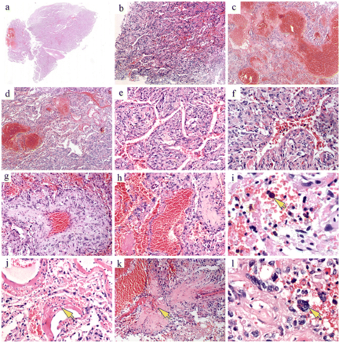
Angiomatous Pleomorphic Xanthoastrocytoma A Case Report And Literature Review Diagnostic Pathology Full Text from media.springernature.com Classically in the temporal lobe in children and young adults. The pxa was added as a separate diagnosis to the 1993 world health organization (who). I see that it has been quite some time since you have posted here, but in case you do get back on, i am wondering what type of chemo you had for pleomorphic xanthoastrocytoma w anaplastic features? Pleomorphic xanthoastrocytoma what do we really know about it? Patients have shorter survival rates when compared to those with who grade ii pleomorphic xanthoastrocytoma. Pxa was first reported by kepes et al,1 and because of its proclivity for the temporal lobes, commonly presents with seizures. Focused pleomorphic xanthoastrocytoma with stained slides of pathology. Pleomorphic xanthoastrocytoma (pxa) and anaplastic pleomorphic xanthoastrocytoma (apxa) are two types of very rare astrocytomas.
Pleomorphic xanthoastrocytoma typically occurs in the cerebral hemisphere (the uppermost sections of the brain) and the leptomeninges, one of the layers covering the brain.
Rare (less than 1% of all astrocytic tumors). Home tumors of the nervous system in children supratentorial tumors in children supratentorial pleomorphic xanthoastrocytomas in children homepage. 2018, article id 6428492, 4 pages, 2018. Case report and literature review. Pleomorphic xanthoastrocytomas affect males and females equally. Composite pleomorphic xanthoastrocytoma and ganglioglioma: Patients have shorter survival rates when compared to those with who grade ii pleomorphic xanthoastrocytoma. Pleomorphic xanthoastrocytoma (pxa) is a rare, benign brain tumor that likely arises from astrocytes, cells in the nervous system that make up the supportive pleomorphic xanthoastrocytoma typically occurs in the cerebral hemisphere, the uppermost sections of the brain and the leptomeninges, one of. Pleomorphic xanthoastrocytoma, abbreviated pxa, is neuropathology tumour classically associated with seizures in children. Anaplastic pleomorphic xanthoastrocytoma is related to pleomorphic xanthoastrocytoma and glioblastoma. Focused pleomorphic xanthoastrocytoma with stained slides of pathology. On histopathological examination, diagnosed as pleomorphic xanthoastrocytoma, of left temporoparietal region. Pleomorphic xanthoastrocytoma what do we really know about it?
This means they begin in the brain or spinal cord pleo. The pleomorphic xanthoastrocytoma (pxa) is a rare tumor type of astrocytic origin that was fi rst described as a distinct neoplastic entity by kepes and coauthors in 1979 16.


Comments
Post a Comment